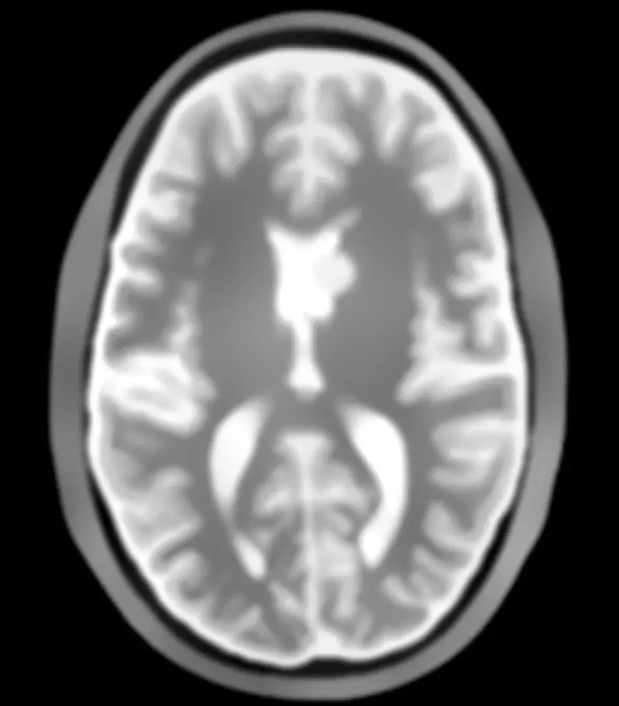
 Tomographic image
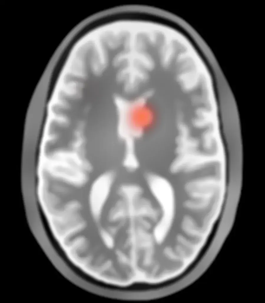 Radar contrast map
How it works
3D brain imaging in seconds, wherever the patient is
01 / 03
Transmit
The device is placed around the patient's head. It emits low-power, non-ionising electromagnetic waves, which propagate into the brain.
02 / 03
Reflect
Waves reflect differently depending on the tissue's dielectric permittivity, a property directly linked to its fluid content. Conditions such as stroke change that balance in the affected area, causing it to stand out from healthy tissue.
03 / 03
Reconstruct
A proprietary AI algorithm solves inverse scattering problems, generating medical images in seconds.
Interoperable by design
Images are generated in DICOM format and are fully interoperable with hospital information systems. Clinicians can view the scans through the traditional viewers they are already used to.
Technology
From leading research to the bedside
Rilemo's device reconstructs brain images from the way body tissues reflect safe electromagnetic waves. The method was developed at the Italian National Institute for Nuclear Physics (INFN) and the European Organisation for Nuclear Research (CERN), and is protected by multiple patents.
Microwaves sit in the same frequency range used every day by mobile networks and Wi-Fi. Unlike CT or MRI, this range requires no heavy shielding, strong magnets or cooling systems, which is what makes it possible to use the device by different operators, in different settings.
Three sequences
Every acquisition produces three sequences at the same time
Tomographic image
Shows the anatomical structure of the head, including the skull, brain parenchyma and main internal compartments, with a resolution comparable to a low-field MRI scan.
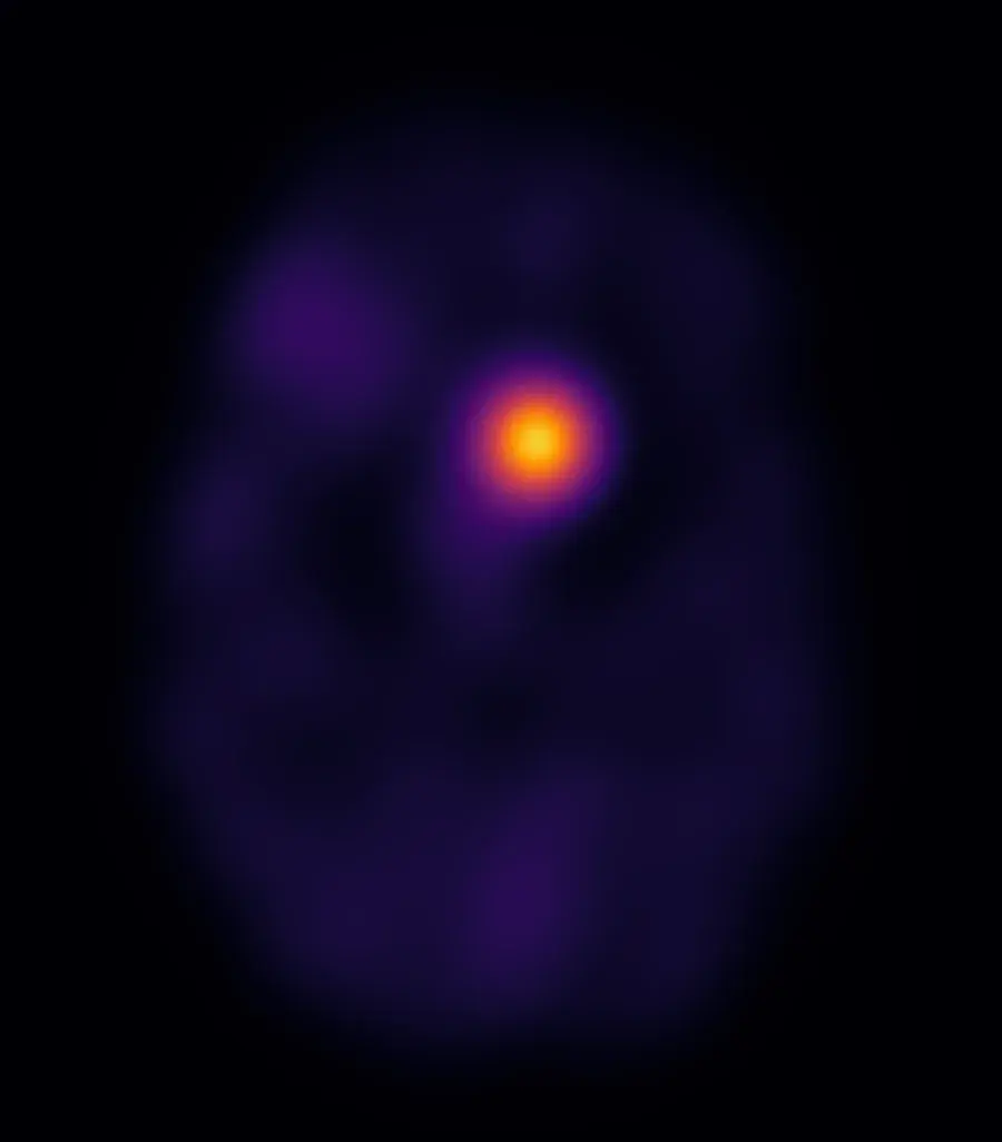
Radar contrast map
Highlights internal echoes and abrupt changes in tissue properties, making boundaries and structural contrast easy to identify.
Dielectric permittivity map
Quantifies the dielectric properties of tissue at every point, showing the homogeneous distribution expected in a healthy head and making deviations easy to spot.
ROADMAP
2020
Research begins
First technology demonstrator built to prove the imaging principle.
Done2025
Ex vivo validation
Acquisition pipeline validated on ex vivo human.
DoneNow
Industrialised · Pre-clinical
Industrialisation completed with a CDMO, ahead of compliance testing and the clinical program.
In progressEarly 2027
In-human clinical trial
Clinical study validating the technology on patients, benchmarked against the gold standard.
PlannedLate 2027 – Early 2028
CE marking · Market release
Certification obtained, opening commercial distribution across European hospital networks.
Planned
Look deeper